

Surgical Microscopes – ToronSMTM™ 1500 Series
Surgical Microscopes – ToronSMTM™ 1500 Series
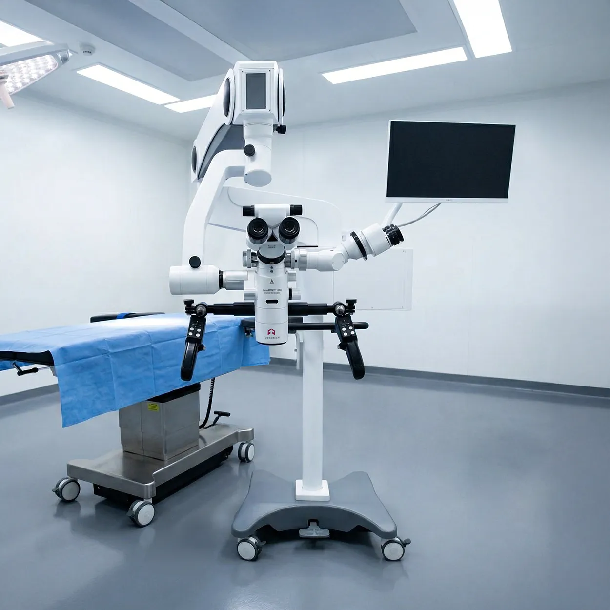
Surgical Microscopes in the ToronSMTM™ 1500 Series are built for operating rooms that need bright coaxial illumination, stable positioning, and fast adjustments during delicate procedures.
This platform combines motorized zoom, motorized working distance, and an ergonomic suspension arm so the microscope can be positioned quickly and held steady. Two xenon light sources and selectable filters help maintain visibility across changing tissue conditions. Integrated imaging and touchscreen control support documentation and team viewing.
ToronSMTM™ 1000 E Series Surgical Microscopes are floor-mounted operating microscopes designed for high-magnification visualization in microsurgery. The system uses continuous, motorized magnification and a VarioDist objective to support a working distance range suitable for common OR setups.
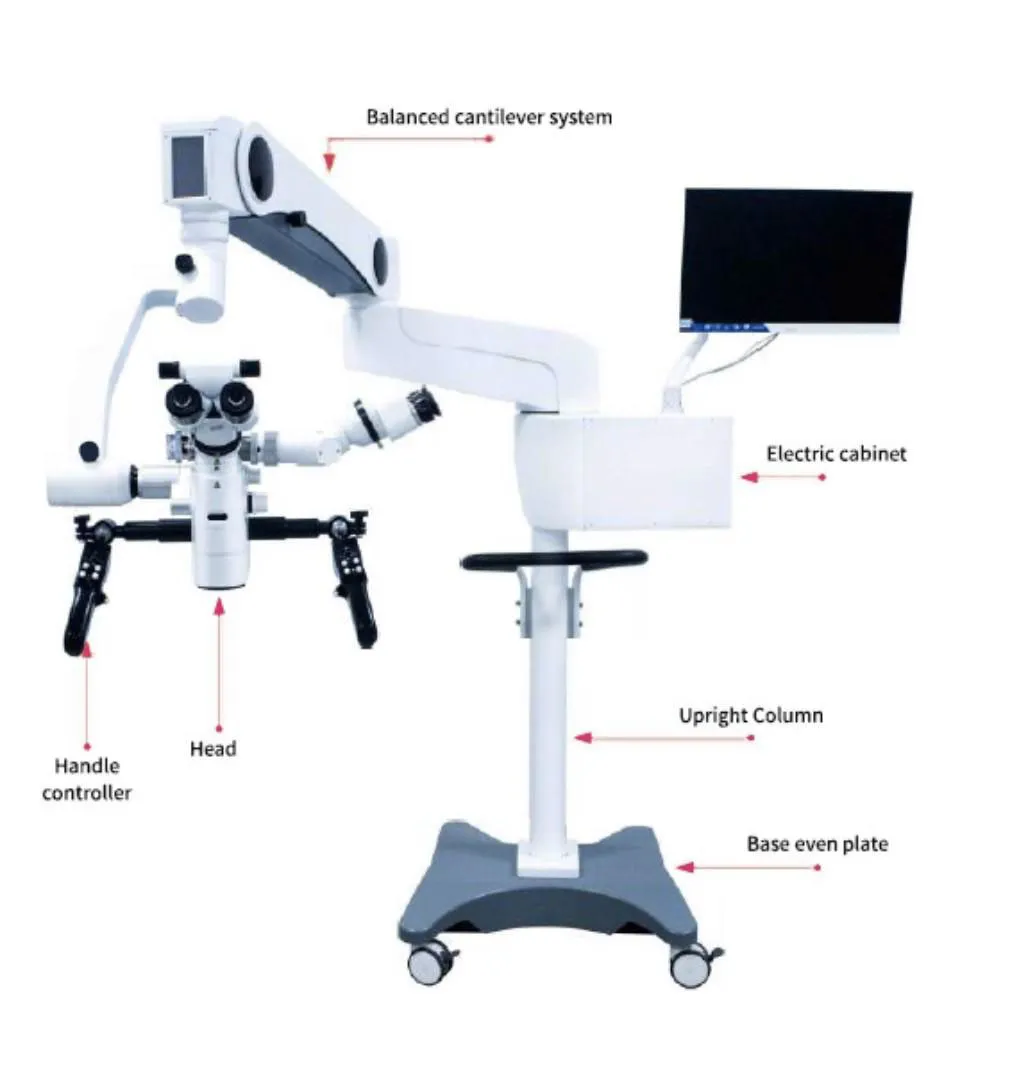
Electromagnetic locking and counter-balanced arm geometry support smooth movement with reliable holding after repositioning. A surgeon handle controller and dual touchscreens streamline control of focus, zoom, illumination, and working distance.
This E Series includes two configurations: ToronSMTM™ 1500 A and ToronSMTM™ 1000 Both share the same core optical and illumination concept, while supporting different stand/arm layouts and installation preferences.
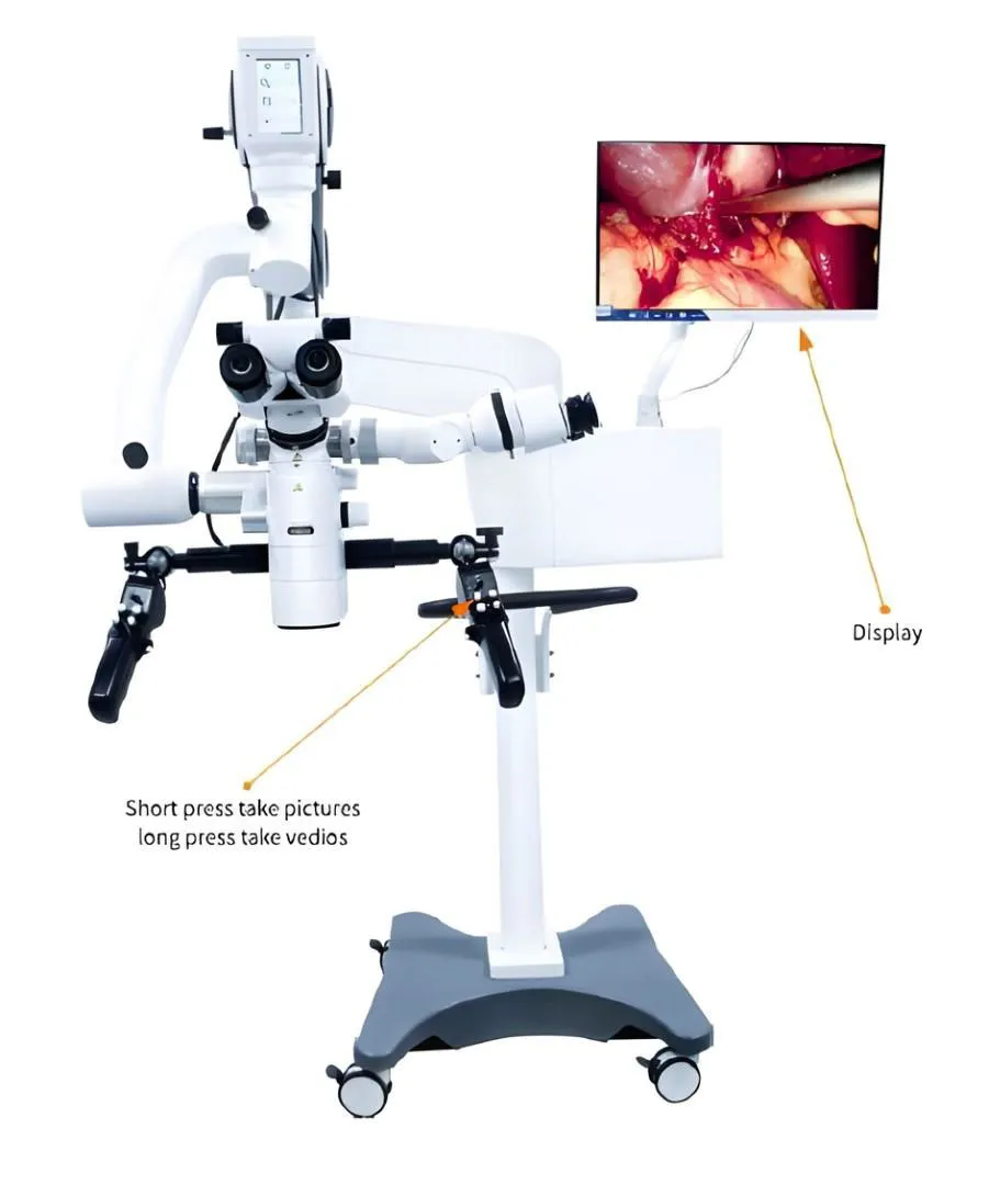
Built-in camera capability and monitor viewing can be used for recording, review, and teaching. The result is a Surgical Microscopes solution that fits neurosurgery and other high-precision disciplines with repeatable setup and consistent imaging.
Surgical Microscopes – ToronSMTM™ 1500 Series Applications
- Neurosurgery: High magnification and bright coaxial lighting help visualize fine anatomical structures and deep surgical fields. Electromagnetic locking supports quick repositioning and stable holding once the target is centered. Optional imaging and monitor viewing support documentation and intra-team communication.
- ENT and otologic procedures: ENT cases often require frequent angle changes and compact working space management. The adjustable binocular system and controlled arm motion help maintain ergonomics during long procedures. Filters and small-spot illumination can help improve contrast for fine dissection steps.
- Spine and orthopedic microsurgery: Spine workflows benefit from stable microscope holding and repeatable positioning as the field shifts between levels. Motorized working distance adjustments help maintain focus without constant manual reconfiguration. The assistant tube options support team viewing when a second observer is required.
- Ophthalmic and plastic/reconstructive work: High-clarity optics and consistent illumination help manage reflective surfaces and delicate tissue planes. A high-point eyepiece design can improve comfort for operators wearing glasses. Photo/video capture supports case records and training needs.
Standards
- ISO 13485 (medical device quality management systems)
Surgical Microscopes – ToronSMTM™ 1500 Series Key Features
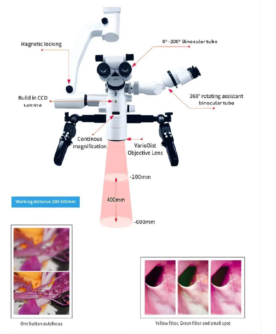
- Continuous motorized zoom with a broad magnification range for smooth transitions from overview to fine detail.
- Motorized working distance (200–600 mm) using a VarioDist objective to support common OR setups.
- Dual xenon illumination system designed to deliver bright lighting with lamp-life tracking shown on-screen.
- Electromagnetic locking + balance system for easy repositioning and stable holding after adjustment.
- Dual touchscreen control for magnification, working distance, and luminance, plus user data storage.
- Surgeon handle controller that supports zoom/focus plus quick photo/video capture (short press photo, long press video).
- Assistant viewing support with rotating assistant tube options and face-to-face viewing configurations for team workflows.
- Built-in imaging workflow (4K option shown) with common output interfaces for recording and display.
Theory and Method
Optical principle in an operating microscope
An operating microscope increases usable detail by combining an objective lens, a zoom system, and eyepieces (or a camera output) to deliver a magnified, high-contrast image. Coaxial illumination sends light along the same axis as the viewing path, which helps reduce shadows in deep fields. Multilayer coatings improve light transmission, while achromatic correction helps reduce color fringing and supports clearer edges at high magnification.
How the ToronSMTM™ 1500 is typically used
The microscope is positioned over the field and balanced so movement feels light and controlled. The operator adjusts magnification, focus, and working distance using the handle controller and touchscreens while maintaining sterile workflow. Electromagnetic locking is released for repositioning and re-engaged to hold the set position. If imaging is enabled, the team can record photos or video and review files for documentation or teaching.
Surgical Microscopes – ToronSMTM™ 1500 Series Technical Specification
| Specification | ToronSMTM™ 1500 A | ToronSMTM™ 1500 B |
|---|---|---|
| Surgical microscope type | Floor stand operating microscope | |
| Eyepieces | 12.5× | |
| Pupil distance adjustment | 55–75 mm | |
| Diopter adjustment | +6D to -6D | |
| Binocular tube | 0°–200° variable angle binocular tube | |
| Magnification / zoom | 6:1 zoom, motorized continuous; system magnification 1.8×–19× | |
| Field of view (main) | Φ12–Φ113 mm | |
| Assistant binocular tube | Face-to-face assistant tube; magnification 3×–16× | |
| Field of view (assistant) | Φ13–Φ94 mm | |
| Illumination | 2 sets xenon lamp; illumination intensity 100,000 lux (each set) | |
| Working distance | Motorized 200–600 mm | |
| Stand / brakes | Electromagnetic brakes | |
| Filters | Yellow filter, green filter, small spot | |
| Maximum arm length | 1715 mm | |
| Height range (objective to floor) | 690 mm ≤ H ≤ 1452 mm | |
| Handle controller functions | 10 functions (zoom, focusing, magnetic lock, photo/video, browse, autofocus, brightness adjust) | |
| Camera system | Built-in camera system; photo/video capture; browse pictures; autofocus function | |
| Weight | 215 kg | |
| Best For | OR teams that need flexible assistant positioning and frequent repositioning during cases | OR teams that prefer a consistent, repeatable surgeon–assistant setup for routine workflows |
| Recommended OR Layout | Rooms where the assistant may switch sides or where space constraints change between procedures | OR layouts where the assistant is typically positioned face-to-face with the primary surgeon for stable collaboration |
| Key Differentiator | Free-rotatable assistant stereoscope supports dynamic assistant placement and angle changes | Face-to-face tube configuration supports a fixed collaboration geometry and predictable ergonomics |
| What stays the same | Same motorized zoom platform, motorized working distance, dual xenon illumination, electromagnetic brakes, filters, handle controls, and imaging workflow | Same motorized zoom platform, motorized working distance, dual xenon illumination, electromagnetic brakes, filters, handle controls, and imaging workflow |
Built-in 4K camera system (as configured in the brochure)
| Camera parameter | Value |
|---|---|
| Image sensor size | 1/1.8 inch |
| Image sensor type | CMOS |
| Scanning method | Progressive scanning |
| Frame rate | 60 fps |
| Effective pixels | 3840(H) × 2160(V) |
| Video coding | H.265 |
| Interfaces (shown) | HDMI 2.0, DVI (DVI25+4), SDI (3G), LVDS (dual 8-bit), RJ45 (1000 Mbit/s), USB 3.0 |
| Minimum illumination | 1 lux (F1.2) |
| Operating temperature | 5°C to 40°C |
| Working humidity | 30% to 85% (non-condensing) |
| Atmospheric pressure | 700 to 1060 hPa |
| Voltage | DC 12V, 2A |
| Power consumption | 7.3 W (12V 604 mA) |
| Camera size (shown) | Φ25 mm × 148 × 100 mm |
Quick buyer guidance
- Choose ToronSMTM™ 1500 A when the assistant’s position changes often or the room layout varies by procedure.
- Choose ToronSMTM™ 1500 B when your team follows a face-to-face workflow and wants consistent ergonomics case after case.