

Surgical Microscopes – ToronSMTM™ 1000 Series
Surgical Microscopes – ToronSMTM™ 1000 Series
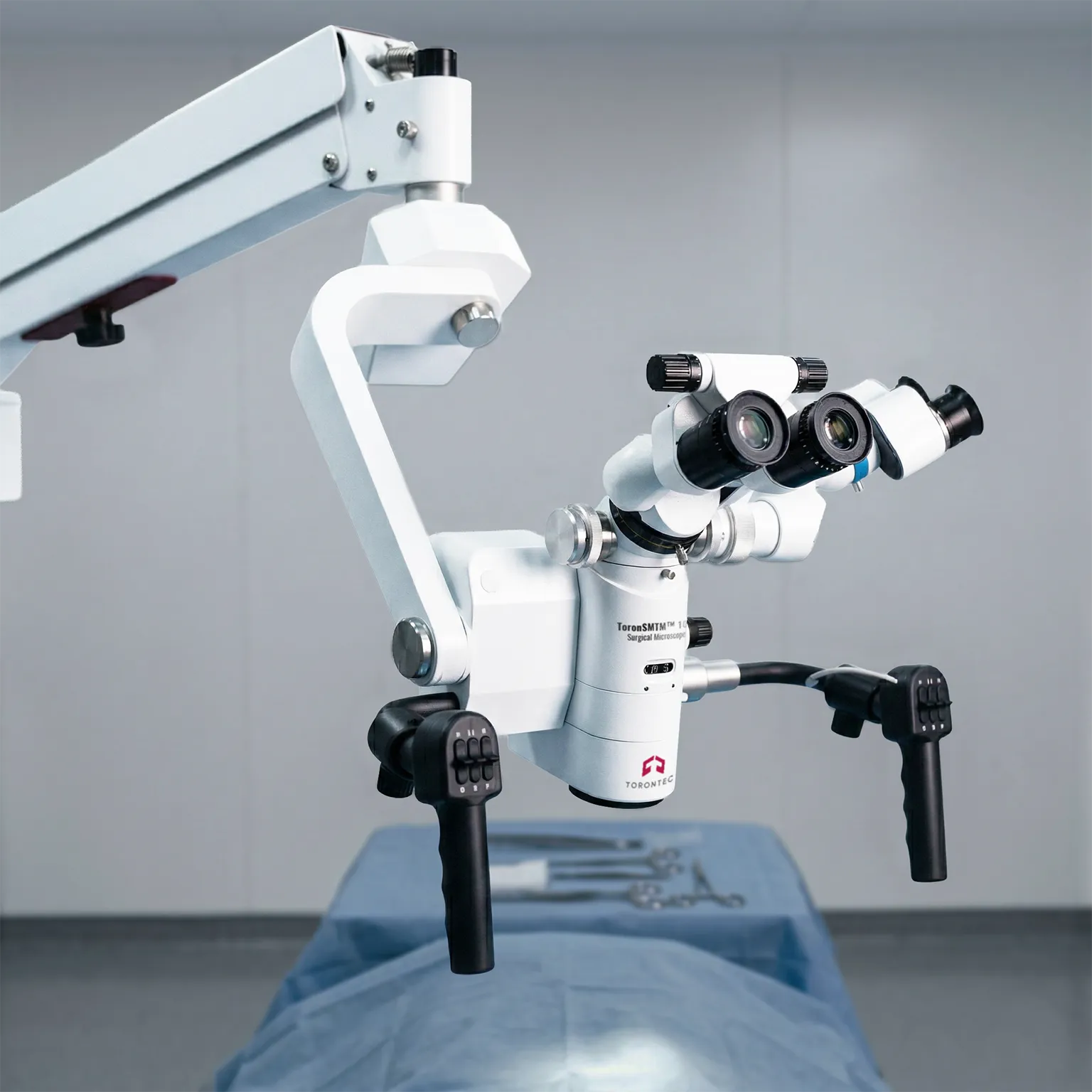
Surgical Microscopes in the ToronSMTM™ 1000 Series support clear, stable visualization for microsurgical workflows. This series is designed for OR environments where consistent illumination, fast focusing, and reliable positioning matter during long procedures. The platform is available in two configurations under the same family, supporting different room layouts and mounting preferences. Options can include integrated video capture and monitor-based viewing for documentation and team visibility.
ToronSMTM™ 1000 Series Surgical Microscopes are optical systems built to magnify fine anatomical detail under bright, even coaxial lighting. They combine motorized zoom with smooth focusing so operators can adjust magnification without breaking workflow. The stand and arm geometry are designed to hold position with controlled movement for ergonomic positioning. Depending on model, the system can support in-procedure photo and video capture with autofocus.
ToronSMTM™ 1000 is focused on compact, high-output LED illumination with a variable working distance objective. Support routine and advanced microsurgical use where clarity, stability, and documentation are required.
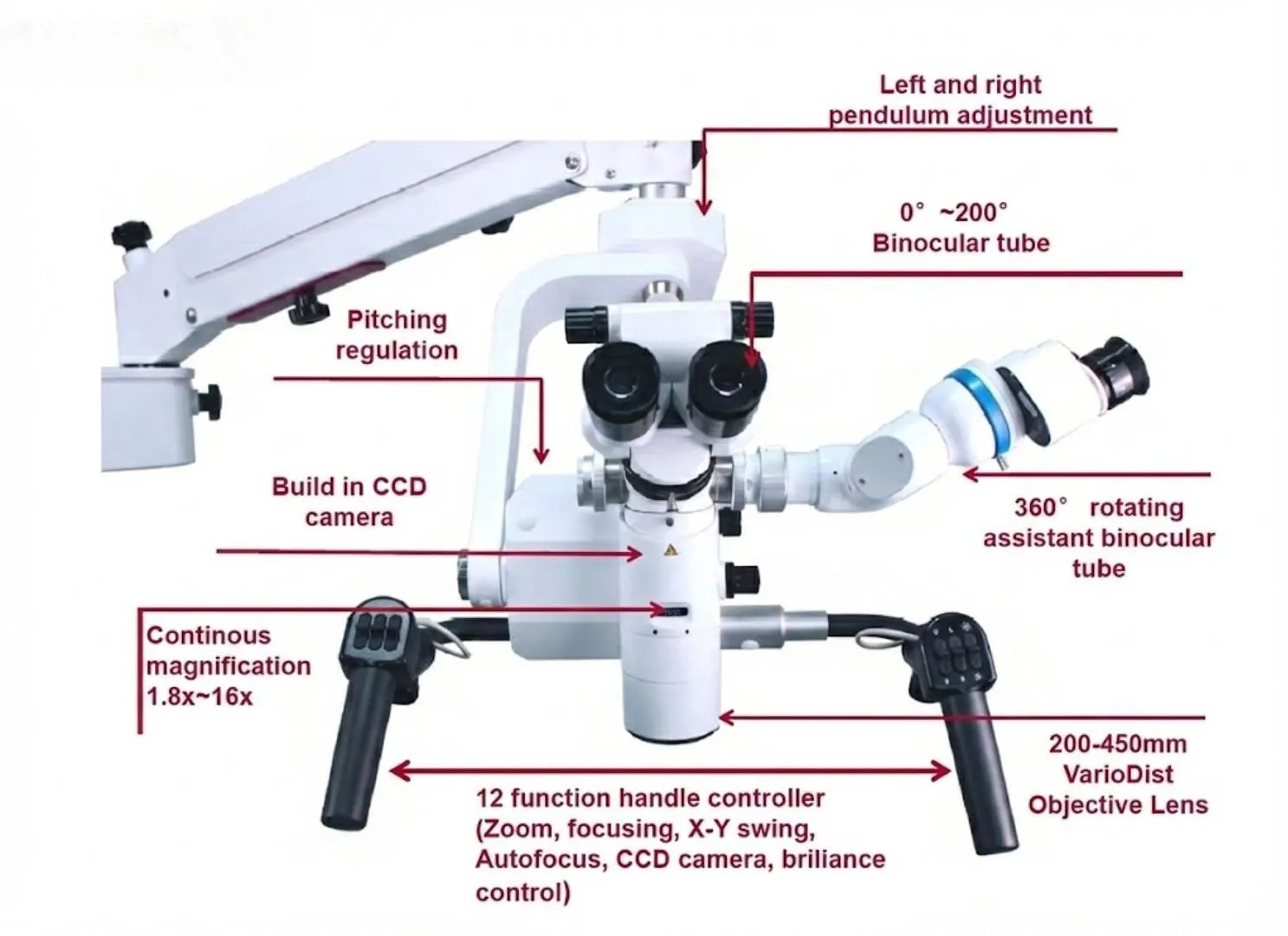
Surgical Microscopes in the ToronSMTM™ 1000 Series support high-magnification visualization during delicate clinical procedures. This series uses a motorized zoom optical system, bright coaxial illumination, and an ergonomic binocular tube to help maintain a stable, comfortable view. Motorized focusing and head movement controls help the operator reposition quickly while keeping the field centered. An assistant binocular tube option supports teaching and collaborative workflows. Camera and autofocus options support documentation needs when required.
Surgical Microscopes – ToronSMTM™ 1000 Series Applications
- Neurosurgery: Fine structures and narrow surgical corridors demand stable illumination and clear depth perception. Motorized zoom and focusing support rapid changes in working depth while keeping image quality consistent. Optional recording supports case review and training.
- ENT: ENT procedures often involve confined spaces and frequent repositioning. The microscope’s head swing and motorized controls help maintain the target in view while adjusting angles. Bright illumination improves contrast when working in deeper cavities.
- Brain Surgery: Precise tissue differentiation requires consistent lighting and accurate magnification control. Continuous zoom supports smooth transitions from overview to detail work without changing posture. Optional autofocus can reduce refocus steps during frequent depth changes.
- Dental / Microsurgery: Dental and microsurgical work benefits from controlled magnification and a comfortable viewing posture during long cases. Variable-angle binocular viewing helps reduce neck strain. Optional imaging supports documentation and patient record requirements.
Standards
- ISO 13485:2016 - Medical Devices – Quality Management System
Surgical Microscopes – ToronSMTM™ 1000 Series Key Features
- Motorized 6:1 continuous zoom for smooth magnification changes during procedures
- High-intensity illumination designed for bright, uniform lighting at the working field
- Variable-angle binocular tube options to support ergonomic posture across long cases
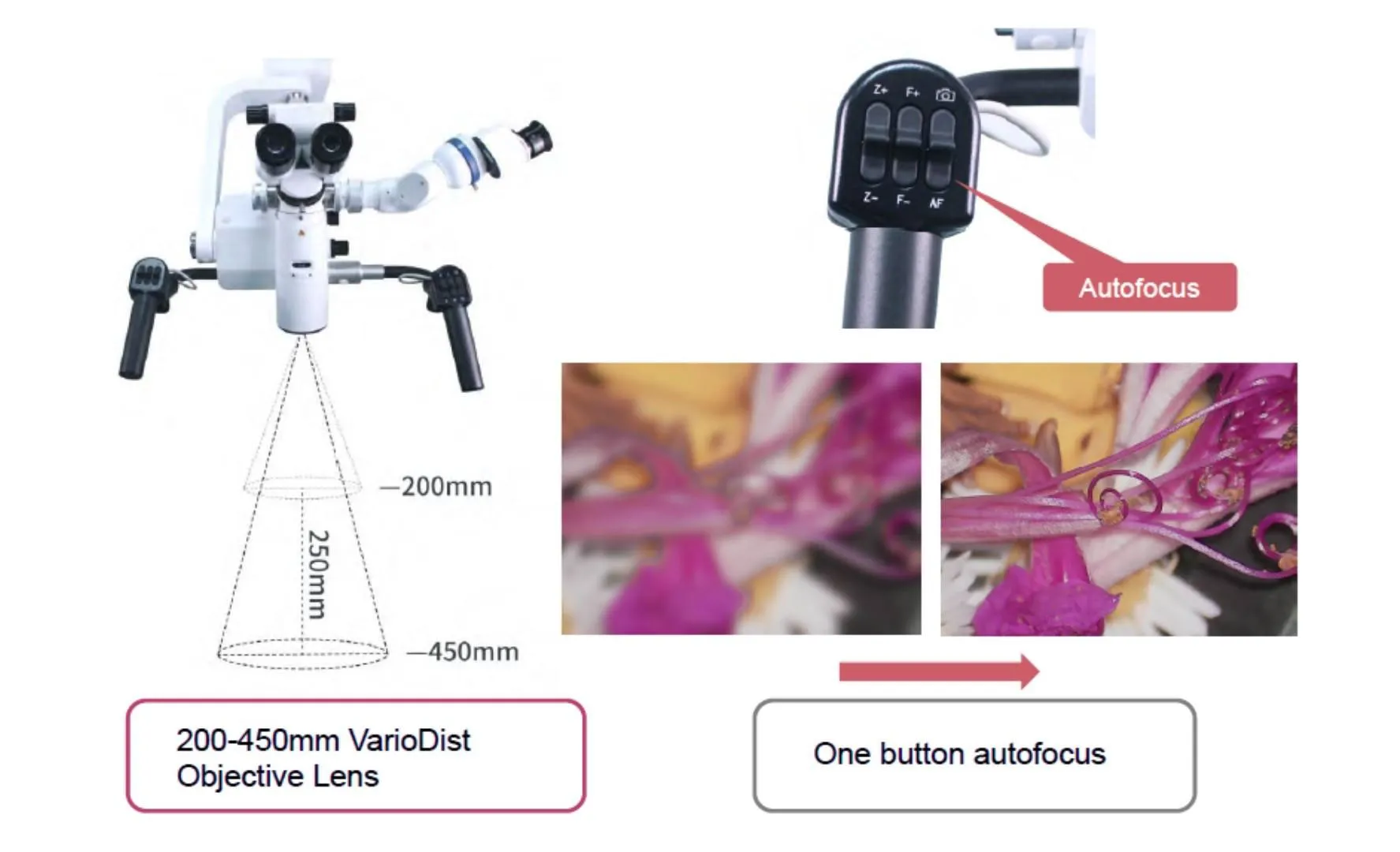
- Motorized focusing to maintain sharpness while changing working depth
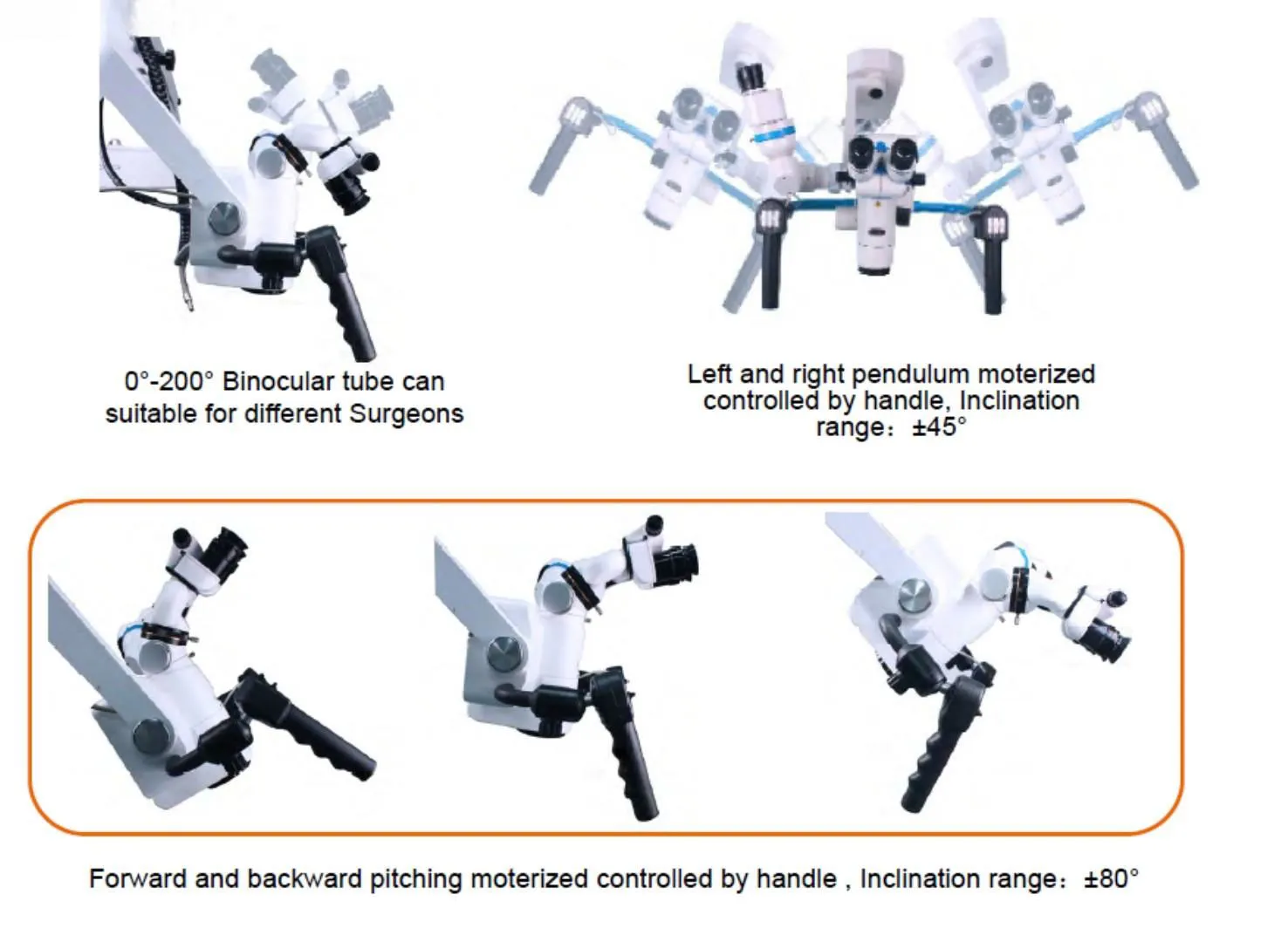
- X–Y head swing control to reposition the view and stop at selected angles
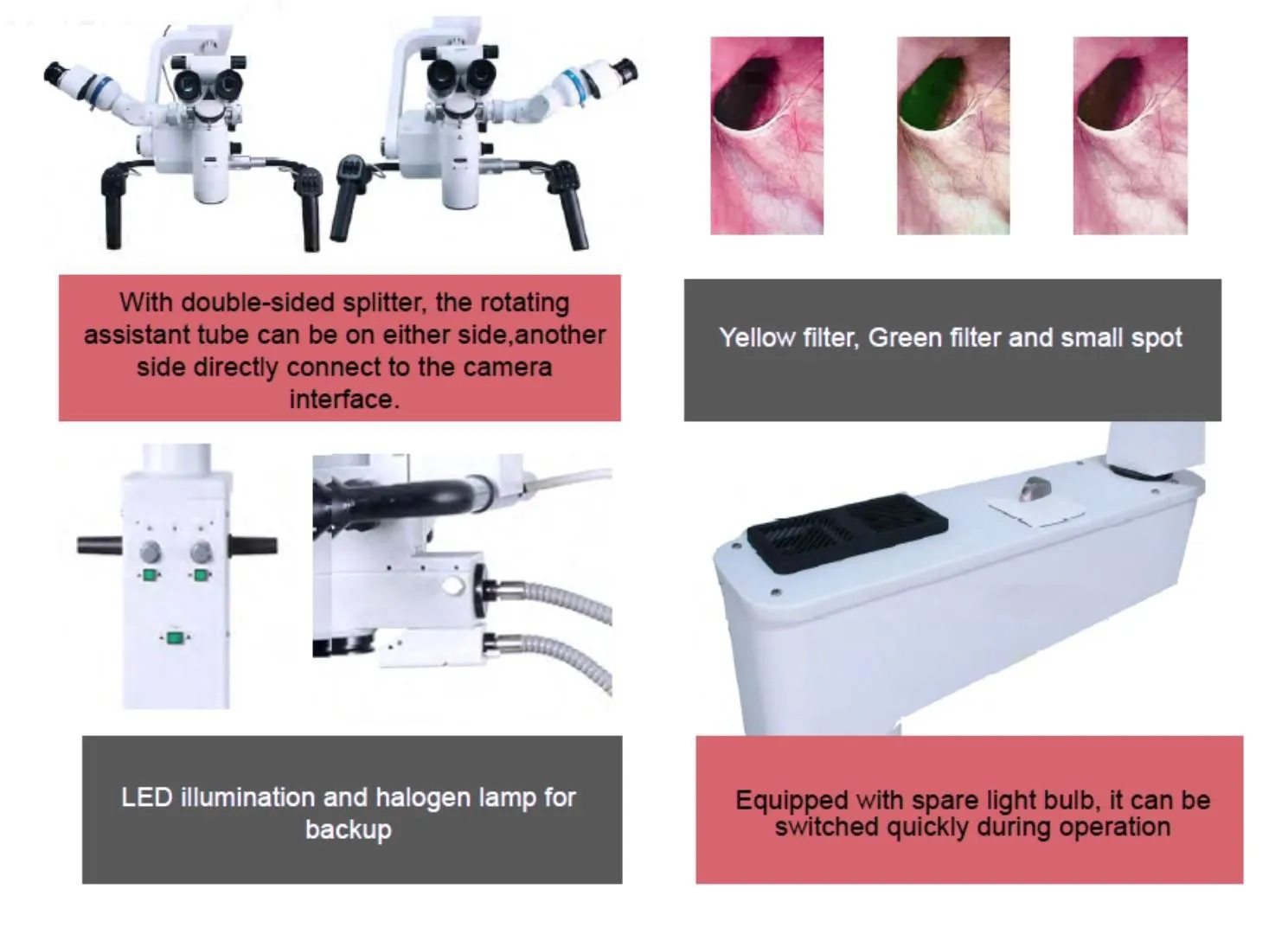
- Assistant binocular tube option for teaching, observation, and teamwork
- Optional autofocus to reduce manual refocusing during frequent depth changes
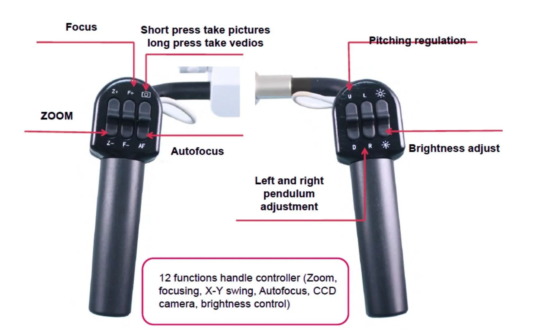
- Optional camera system for photo/video capture and image browsing via handle control
Theory and Method
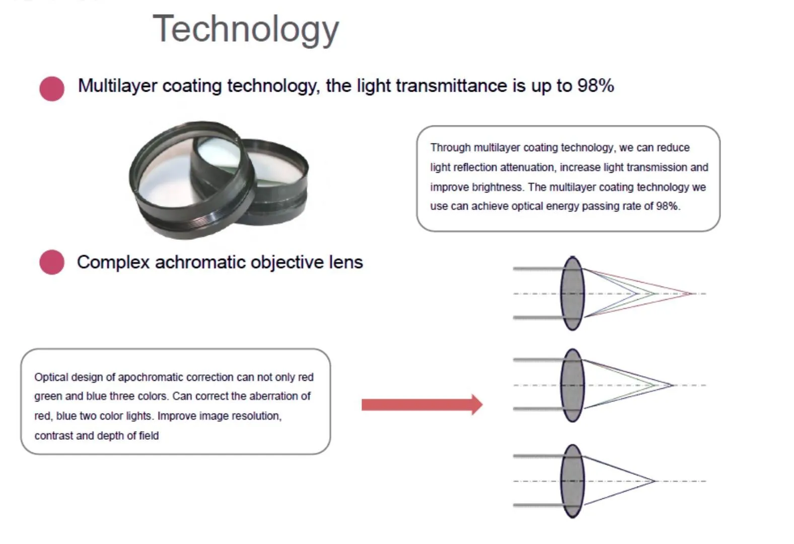
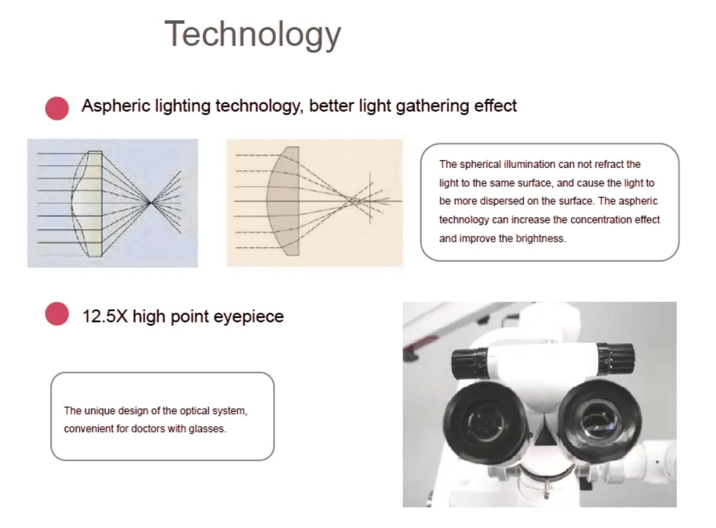
A Surgical Microscope forms a magnified image of the surgical field using an objective lens, zoom optics, and eyepieces. Coaxial illumination directs light along the same axis as observation, improving visibility in deep or narrow operative sites.
The operator selects a magnification level with continuous zoom, then uses motorized focusing to lock sharpness at the working distance. Head swing controls help reposition the view in X and Y directions without losing alignment. When equipped, an imaging module captures photos or video for documentation and training.
Surgical Microscopes – ToronSMTM™ 1000 Series Technical Specification
| Specification | ToronSMTM™ 1000 |
|---|---|
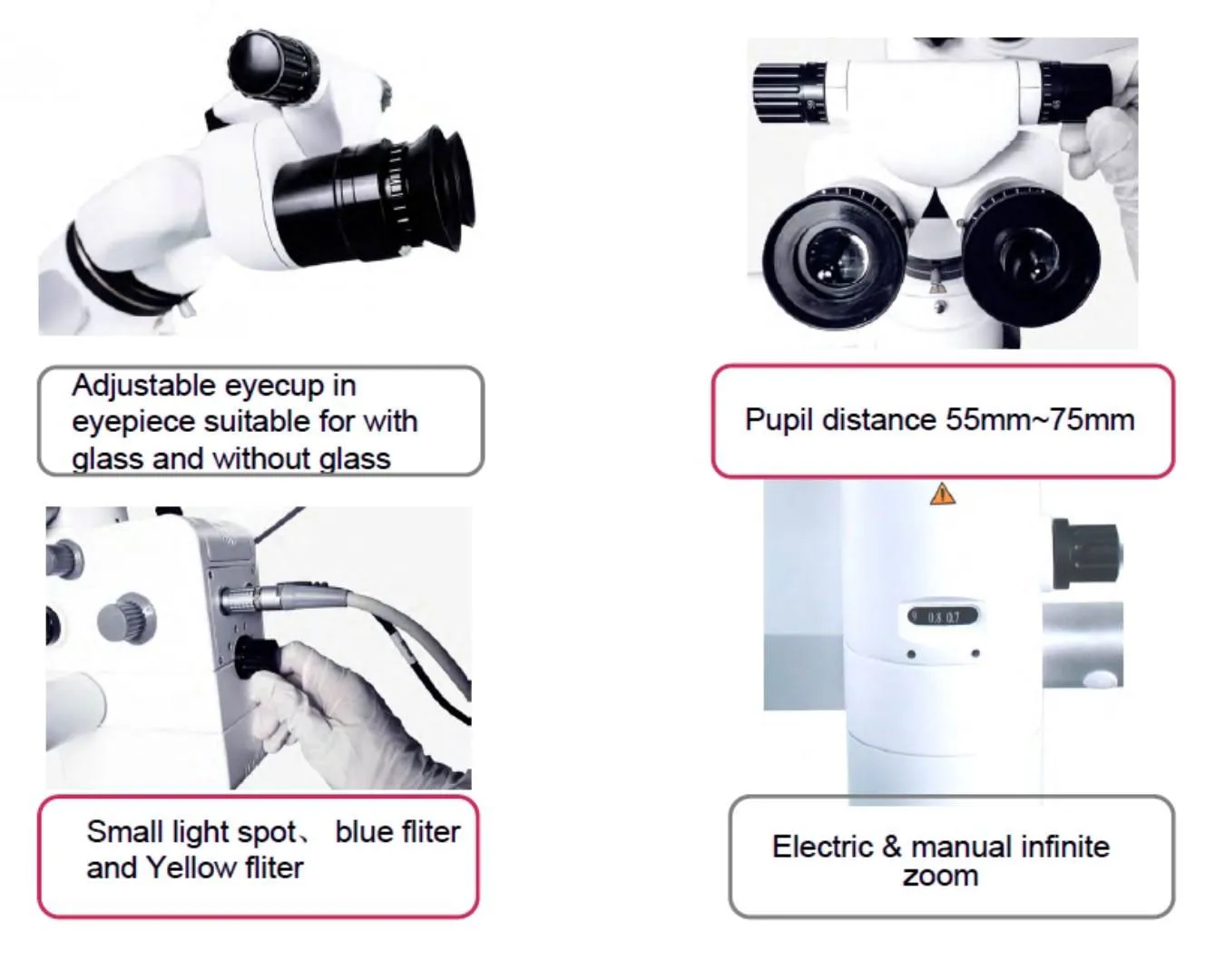
| Eyepieces | 12.5×; pupil distance 55–75 mm; diopter +6D to –6D |
| Binocular tube | 0°–200° variable angle; added pupil distance adjustment |
| Zoom / magnification | 6:1 zoom, motorized continuous; 1.8×–21× |
| Field of view | Φ13–Φ110 mm |
| Assistant binocular tube | 360° rotating; 3×–16×; FOV Φ74–Φ12 mm |
| Illumination | 100 W LED; lifetime >100,000 hours; up to 100,000 lux |
| Objective / working distance | Motorized 200–450 mm (VarioDist style) |
| Focusing | Motorized 200–450 mm |
| X–Y swing | X: ±45° motorized; Y: +90°; stops at any angle |
| Filters | Yellow filter, green filter, small spot |
| Max arm length | 1200 mm |
| Stand / height range | Carrier arm swing 0–300°; objective-to-floor 800 mm < H < 1250 mm |
| Handle controller | 12 functions (zoom, focus, X–Y swing, photo/video, browse images, autofocus, brightness adjust) |
| Camera system | Optional built-in 4K camera; photo/video; image browsing |
| Autofocus | Optional |
| Weight | 169 kg |
| Typical departments | Neurosurgery, ENT, brain surgery, dental |