
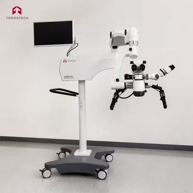
4K Surgical Microscope
4K Surgical Microscope - ToronSM™ 640
Operating Microscope with Integrated 4K Video for Neurosurgery, ENT, and Spine
ToronSM™ 640 4K surgical microscope gives operating teams a stable, high-magnification view of the surgical field with 4K video capture built directly into the optical head. Motorized zoom across a 1:6 ratio and a VarioDist objective covering 200 mm to 630 mm let the surgeon change magnification and working distance without breaking sterile position. Dual 300 W xenon sources deliver coaxial illumination deep into narrow corridors. The system is configured for neurosurgery, ENT, and spine procedures.
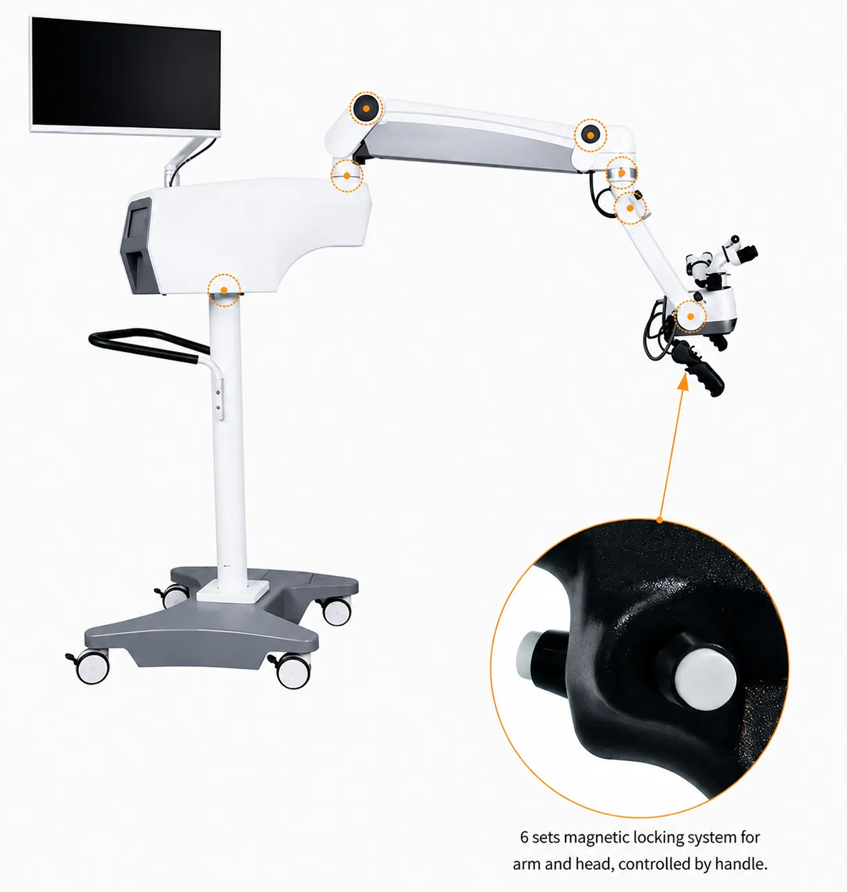
Six electromagnetic brakes hold every joint of the balanced cantilever arm and release together from either handle, so repositioning takes one button press and one smooth movement. A 360° rotating assistant tube, an inclinable main binocular tube, and adjustable eyecups let two surgeons work at different postures on the same field. Two programmable touch screens and programmable handles store settings for up to ten users.
Hospitals adopting the ToronSM™ 640 gain a documentation-ready operating microscope. The integrated 4K CMOS camera records to USB storage and outputs simultaneously over HDMI, DVI, SDI, LVDS, and RJ45 for wall displays, teaching rooms, and surgical video archives. Optional near-infrared fluorescence hardware extends the system to ICG vascular imaging.
4K Surgical Microscope - ToronSM™ 640 Applications
The ToronSM™ 640 supports microsurgical disciplines that depend on high optical resolution, long working distance, and shadow-free coaxial light.
- Neurosurgery: magnified visualization of cortical and subcortical anatomy during tumor resection, aneurysm clipping, and arteriovenous malformation treatment
- Cerebrovascular Surgery: intraoperative assessment of vessel patency during cerebral artery bypass, aided by optional ICG fluorescence imaging
- Spine Surgery: illumination and magnification of the canal and neural elements during decompression, discectomy, and fusion procedures through narrow retractor corridors
- ENT and Otology: middle ear, mastoid, and skull base work where the 200 mm to 630 mm working distance range accommodates deep, angled approaches
- Epilepsy Surgery: precise identification of resection margins in functionally sensitive cortex
- Ophthalmic Surgery: anterior and posterior segment procedures requiring fine focus control and adjustable illumination spot size
- Orthopedic and Reconstructive Microsurgery: nerve repair, vessel anastomosis, and free flap procedures that call for stereoscopic depth perception at high magnification
- Surgical Training and Telemedicine: 4K video output and dual-observer optics for resident teaching, case review, and remote consultation
4K Surgical Microscope - ToronSM™ 640 Key Features
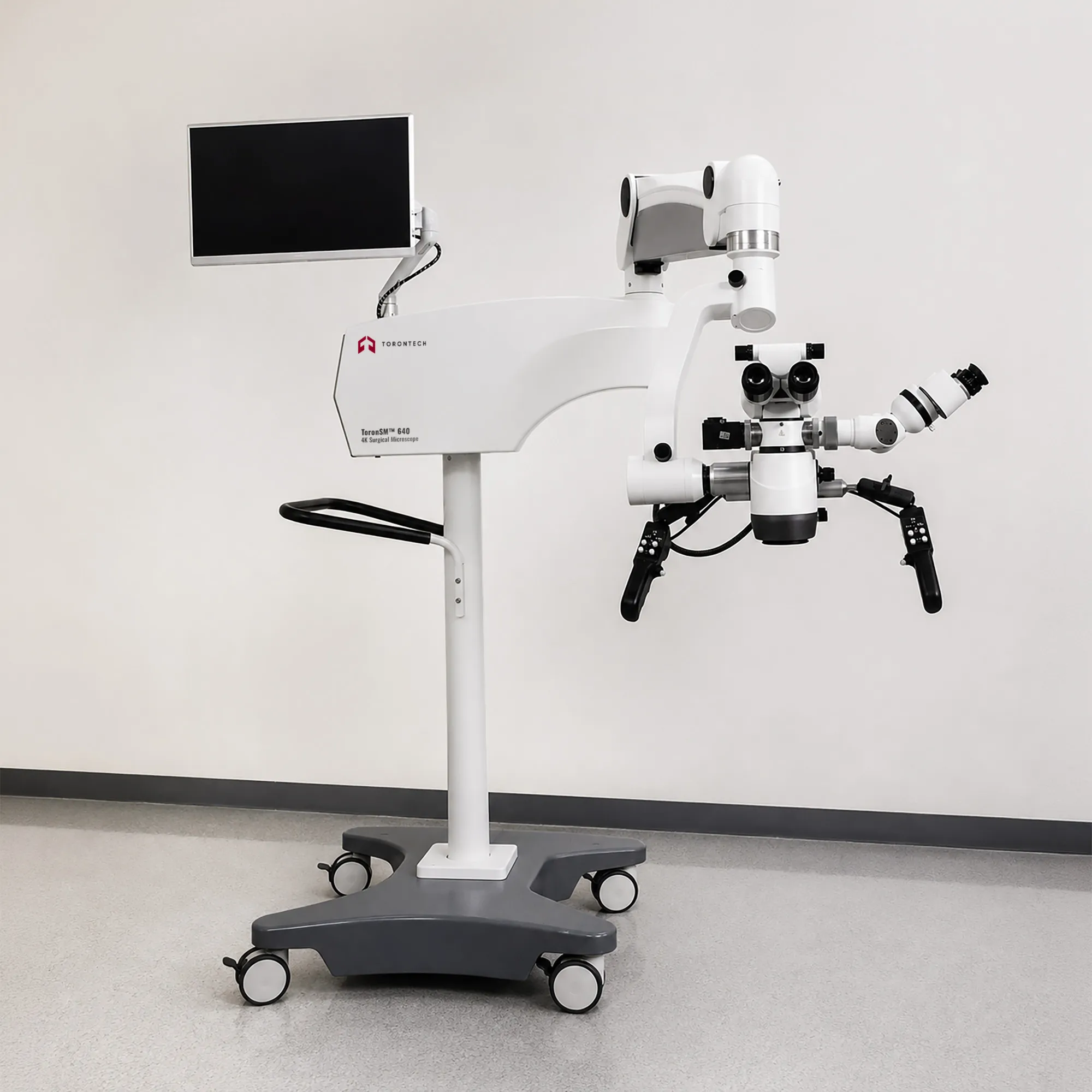
The ToronSM™ 640 combines motorized optics, dual-source illumination, and integrated video in one floor-stand platform.
- Six-Point Electromagnetic Braking: all arm and head joints lock and release together from a handle button, giving one-handed repositioning and a firm hold once released
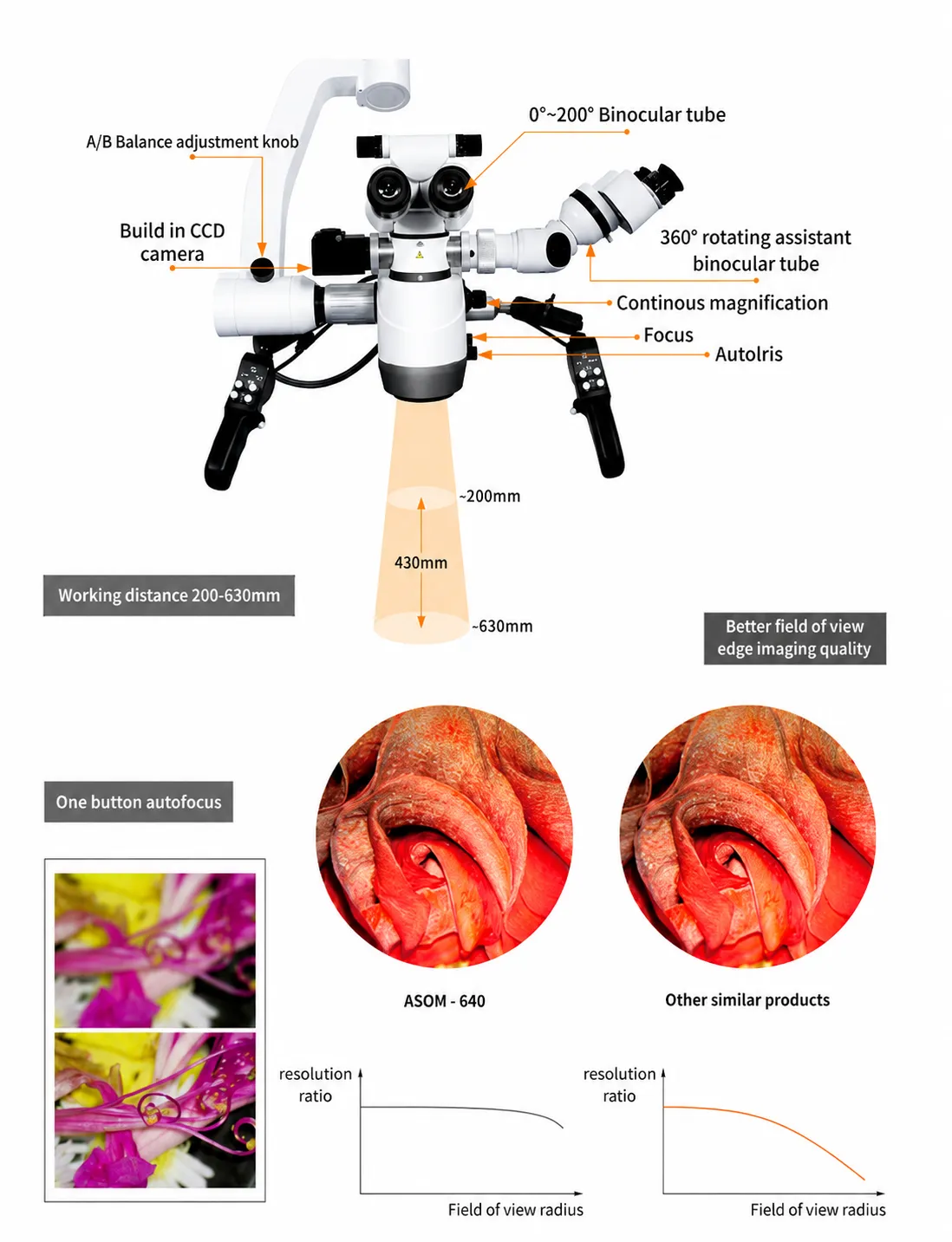
- Motorized 1:6 Zoom with Autofocus: single-button focus and continuous magnification control keep the surgeon's hands on the handles and eyes on the field
- 200 mm to 630 mm VarioDist Objective: electronic focus across the full working distance range removes the need to change objective lenses between procedures
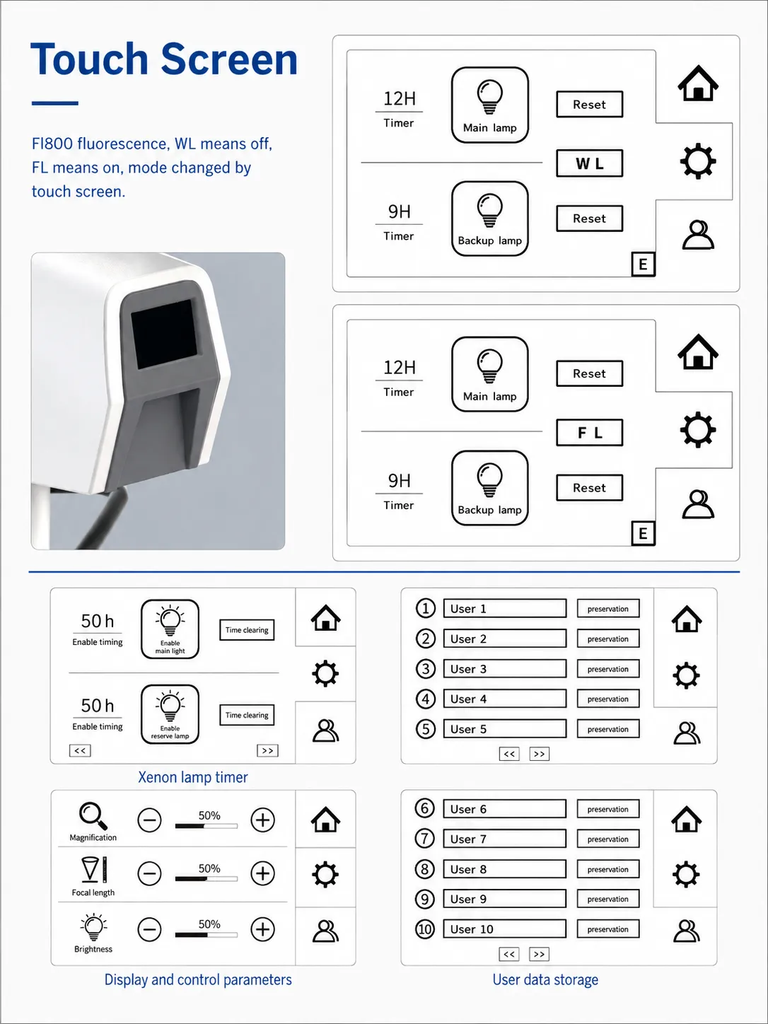
- Dual 300 W Xenon Illumination: a main source plus an on-board backup source, both with lamp-hour counters on the touch screen, reduce the risk of an intraoperative light failure
- Automatic and Manual Spot Tracking: illumination spot size follows magnification in automatic mode or adjusts manually down to an 11 mm minimum for narrow corridors
- Integrated 4K Video with 60 fps Capture: a 1/1.8-inch CMOS sensor records stills and video straight to USB storage and feeds HDMI, DVI, SDI, LVDS, and RJ45 outputs at once
- 0° to 200° Inclinable Binocular Tube: adjustable tilt with 48 mm to 78 mm interpupillary range and adjustable eyecups suits surgeons with or without eyeglasses
- 360° Rotating Assistant Tube: a double-sided splitter places the second observer at 90° or 180° to the primary surgeon, with the opposite port free for the camera
- 12.5x High-Eyepoint Eyepieces: extended eye relief keeps the full field visible for surgeons wearing glasses
- Dual Programmable Touch Screens: 7-inch panels display lamp timers, magnification, focal length, and brightness, and store presets for up to ten named users
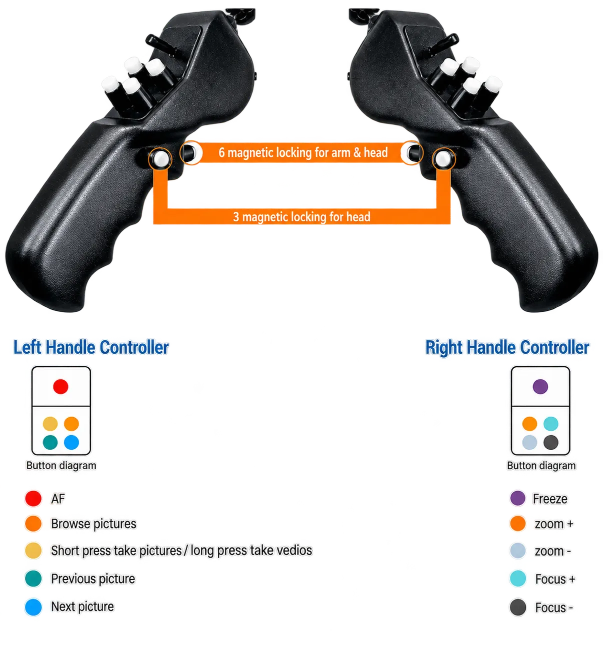
- Programmable Handle Controllers: left and right grips assign zoom, focus, brake release, image capture, video recording, playback, and brightness to individual buttons
- Optional Programmable Footswitch: duplicates zoom, focus, capture, autofocus, and brightness control for hands-free operation
- Optional Fluorescence Module: 800 nm infrared vascular fluorescence hardware supports ICG imaging, including a fusion display that overlays fluorescence on the live video
- Balanced Cantilever with Electronic Damping: manual balancing on all axes plus damping holds the head steady at high magnification
- X-Y Translator: 80 mm by 80 mm fine positioning with a centering button for precise field adjustment
- Laser-Compatible Design: the head accepts surgical laser use, with a focus lock that maintains alignment when a laser micromanipulator is fitted
Theory and Method
Surgical microscopes create depth perception by splitting light from a single large objective lens into two separate optical channels, one for each eye. The two channels sample the beam at slightly different angles, and the brain fuses the resulting pair of images into a three-dimensional view. Magnification changes through a Galilean zoom system, a set of movable lens groups that alter the beam path length without shifting focus. The ToronSM™ 640 motorizes that zoom system across a 1:6 range and pairs it with an apochromatic objective that corrects red, green, and blue focus points to a common plane.
Working distance equals the focal length of the objective, so the VarioDist lens changes focus electronically from 200 mm to 630 mm rather than through a swapped lens. Multilayer antireflection coatings raise transmittance to 98% by suppressing the reflections that occur at each glass-to-air surface, which matters across the many elements in a stereo microscope. Light reaches the field coaxially through fiber optics and an aspheric condenser, which converges rays to a tighter, brighter spot than a spherical lens can.
Aperture size governs the trade-off between brightness and depth of field. Opening the iris admits more light but shortens the range of sharp focus, while closing it extends depth at the cost of brightness. The AutoIris function ties aperture and spot diameter to the zoom position, so the illuminated area tracks the field of view automatically and the operator avoids overfilling the wound with light. Near-infrared fluorescence imaging exploits indocyanine green, which absorbs light near 800 nm and re-emits at a longer wavelength that a filtered camera isolates from the visible surgical image.
4K Surgical Microscope - ToronSM™ 640 Technical Specifications
| Parameter | Specification |
|---|---|
| Model | ToronSM™ 640 |
| Application | Neurosurgery, ENT, spine |
| Magnification | Motorized zoom system, 1:6 zoom ratio |
| Working Distance | 200 mm to 630 mm, motorized multifocal lens, continuously adjustable |
| Binocular Tube | 0° to 200° inclinable |
| Eyepiece | 12.5x / 10x |
| Interpupillary Distance | 48 mm to 78 mm |
| Diopter Adjustment | +7 D to -7 D or +5 D to -8 D |
| Field of View | F13.2 mm to F180 mm |
| Brake System (Head and Arm) | 6 sets, electromagnetic |
| Multifunctional Programmable Handles | Motorized zoom +/-, motorized focus +/-, lock and unlock 3 magnetic brakes, lock and unlock 6 magnetic brakes, video recording, photo capture, image browsing, autofocus, brightness +/-, X-Y movement |
| Touch Screen | 7-inch programmable, displays operating status and main/backup illumination indicators, user profile setting and storage |
| Assistant Tube | 360° rotating |
| Maximum Resolution | 120 line pairs |
| Illumination System | Coaxial |
| Light Source | 300 W main source and 300 W emergency source |
| Lamp Switching | Controlled from touch screen |
| Light Intensity Adjustment | 30% automatic intensity increase at maximum magnification; continuous adjustment 0 to 150,000 lux; working-distance dependent brightness limitation; automatic synchronization of light field diameter with magnification |
| Field of Illumination | Greater than F90 mm |
| Minimum Spot Size | 11 mm |
| Mount | Floor stand |
| Maximum Arm Extension | 1,980 mm |
| Height from Objective to Floor | 690 mm to 1,452 mm |
| Cantilever Height Adjustment Range | 1,400 mm to 2,240 mm |
| Minimum Height of Bracket | 1,850 mm |
| Cross Arm Rotation Angle | 330° |
| Cantilever Rotation Angle | 330° |
| Base Size | 742 mm × 826 mm |
| Base Brake System | Four-wheel brake |
| Optical Transmittance | Up to 98% (multilayer coating) |
Integrated Video System
| Parameter | Specification |
|---|---|
| Image Sensor Size | 1/1.8 inch |
| Image Sensor Type | CMOS |
| Scanning Method | Progressive |
| Frame Rate | 60 fps |
| Effective Pixels | 3,840 (H) × 2,160 (V) |
| Horizontal Resolution | ≥ 1,700 lines |
| Vertical Resolution | ≥ 1,200 lines |
| Luminance Response | Linear fitting coefficient R² ≥ 0.99 |
| Signal to Noise Ratio | ≥ 50 dB |
| Spatial Frequency Response | MTF50: 38 C/°, MTF30: 43 C/° |
| Static Image Tolerance | ≥ 150 |
| Minimum Illumination | 1 lux at F1.2 |
| Video Coding | H.265 |
| Output Modes | HDMI 2.0, DVI 25+4, SDI 3G, dual 8-bit LVDS, RJ45 1000 Mbit/s, USB 3.0 |
| Storage | Automatic recording to USB drive |
| Voltage | 12 V DC, 2 A |
| Power Consumption | 7.3 W (12 V, 604 mA) |
| Operating Temperature | 5 °C to 40 °C |
| Working Humidity | 30% to 85%, non-condensing |
| Atmospheric Pressure | 700 hPa to 1,060 hPa |
| Camera Dimensions | Ø25 mm, 148 mm × 100 mm |
Accessories and Options
| Item | Description |
|---|---|
| Fluorescence Module | 800 nm infrared vascular fluorescence module |
| Programmable Footswitch | Motorized zoom +/-, motorized focus +/-, video recording, photo capture, image browsing, autofocus, brightness +/- |
| X-Y Translator | 80 mm × 80 mm travel with centering button |
| Video Adapter | Sony or Canon camera mount |
| Three-Person Six-Eye Head | Optional microscope head configuration for a third observer |
| Camera-Assisted Focus | Motorized focus with optional camera-assisted adjustment |
| Quality Control Software | Integrated software for quality control and traceability |
Additional Capabilities
| Capability | Description |
|---|---|
| Laser Compatibility | Compatible with surgical laser use |
| Laser Focus Lock | Safety mechanism locks focusing during laser micromanipulator use to maintain alignment |
| Axis Balancing | Manual balancing on all axes |
| Image Stability | Integrated electronic damping system |
| Packaging | Main body box with rack, separate head package box, attachment package box, upper and lower metal plates |