
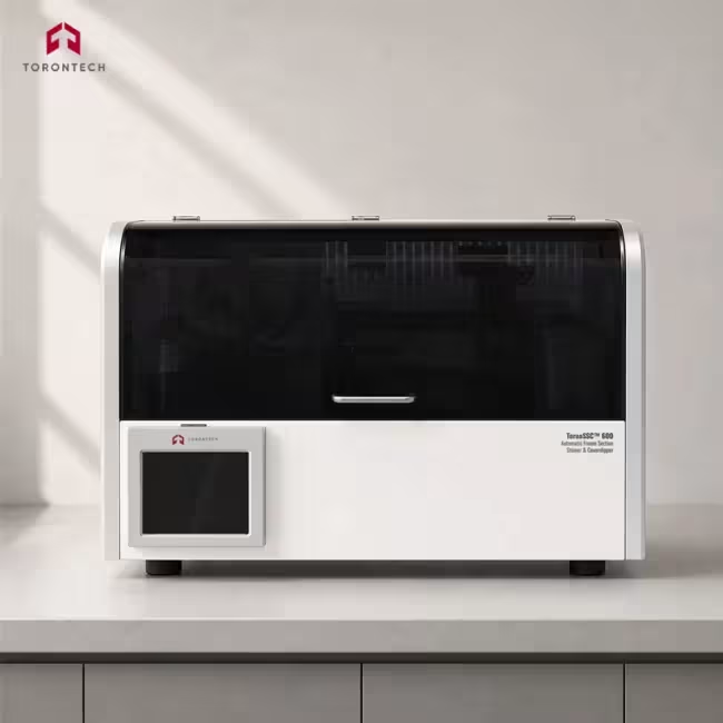
Automatic Frozen Section Stainer & Coverslipper
Automatic Frozen Section Stainer & Coverslipper - ToronSSC™ 600
ToronSSC™ 600 Automatic Frozen Section Stainer & Coverslipper completes staining and glass coverslipping in four minutes, built around the turnaround pressure of intraoperative diagnosis. Torontech supplies this unit to hospital pathology departments where a surgeon waits on the result while the patient remains under anesthesia. The instrument holds 24 stations in a cabinet under 1 m long, covers at 800 slides per hour, and also runs routine tissue H&E and Papanicolaou methods. Covering starts on its own once staining ends.
Three working modes let the instrument match the task at hand. Staining and covering run as one sequence for finished slides, or the operator selects staining alone or covering alone when only part of the workflow is needed. That flexibility lets the unit serve as a standalone coverslipper for an existing stainer during quieter periods. Program memory holds 200 methods of up to 100 steps each, so frozen, routine, and cytology protocols stay loaded together.
An adaptive staining control system adjusts programs against reagent condition, and a reagent management function tracks days in service and basket counts before prompting replacement. A fully enclosed cabinet with activated carbon adsorption and exhaust extraction handles xylene and alcohol vapor at source. Slides can be added at any point during a run, so a second specimen from the same operation enters without a wait. Water valves open only when the protocol calls for them.
Automatic Frozen Section Stainer & Coverslipper - ToronSSC™ 600 Applications
The ToronSSC™ 600 serves laboratories where frozen section turnaround drives the workflow, while still covering the routine staining a pathology department runs the rest of the day.
- Intraoperative Frozen Section Consultation: rapid H&E on cryostat sections so the pathologist can report to the operating room inside the 20-minute window most departments work to
- Margin Assessment in Oncologic Surgery: fast, repeatable staining of resection margins where the surgeon decides how much further to excise before closing
- Mohs Micrographic Surgery: stage-by-stage staining of frozen tissue layers in dermatologic surgery, where each cycle keeps the patient waiting on the result
- Routine Surgical Histology: standard H&E on paraffin sections between frozen cases, using the same instrument rather than a second stainer on the bench
- Cytology and Papanicolaou Screening: polychrome Pap staining held in stored programs, giving consistent nuclear and cytoplasmic contrast for screening work
- Operating Room Adjacent Laboratories: a sub-1 m footprint fits satellite frozen section rooms and cramped suites near the surgical floor where a full-size stainer will not go
- Digital Pathology and Telepathology: uniform mountant film with no overflow or bubbles produces clean scans for remote intraoperative consultation and slide archiving
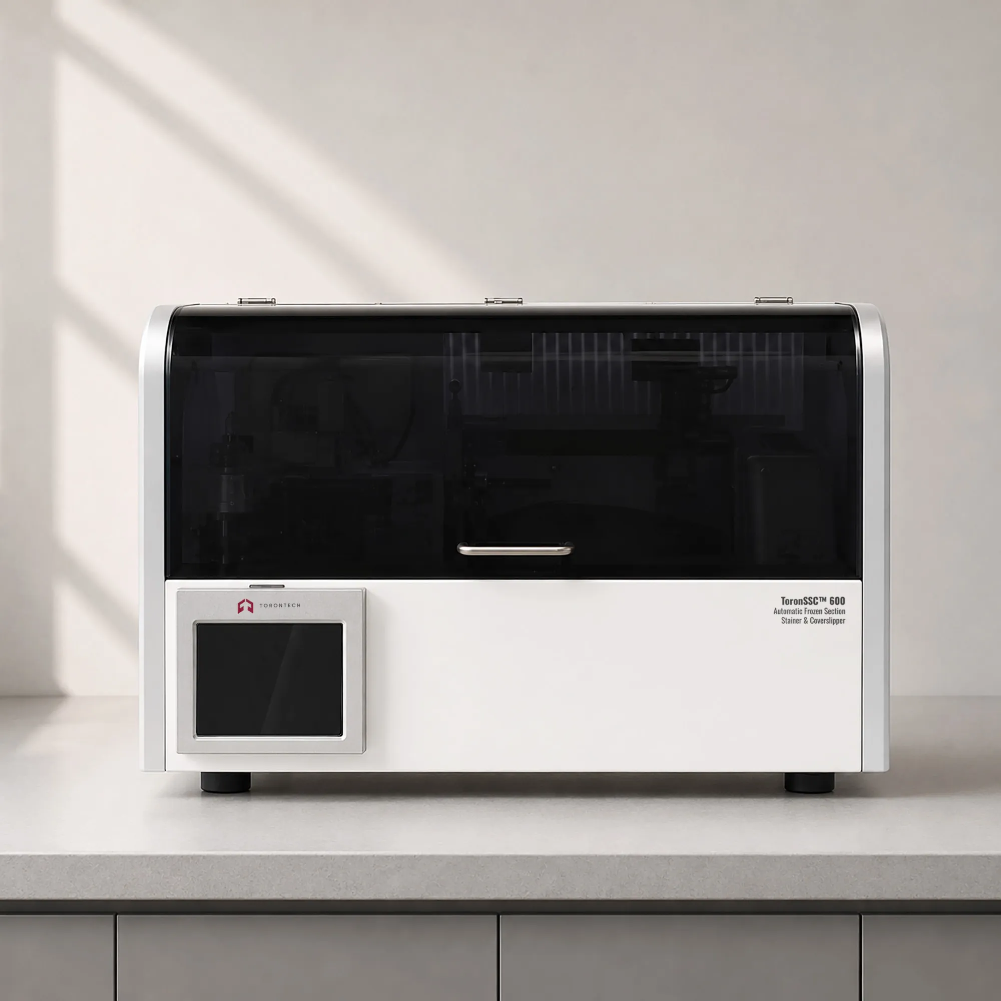
Automatic Frozen Section Stainer & Coverslipper - ToronSSC™ 600 Key Features
The ToronSSC™ 600 pairs a four-minute frozen section cycle with automatic coverslipping and mode switching, in a cabinet small enough for a satellite frozen section room.
- Four-Minute Stain and Cover Cycle: the full frozen section sequence finishes in four minutes, keeping the histology step from becoming the bottleneck in intraoperative reporting
- Three Working Modes: staining with covering, staining only, or covering only, so the instrument adapts to whichever part of the workflow the laboratory needs at that moment
- Standalone Coverslipper Capability: covering-only mode lets the unit finish slides from a separate stainer, adding capacity without a dedicated second machine
- Multi-Protocol Support: rapid frozen, routine tissue H&E, and Papanicolaou methods all run on the same platform, so one instrument covers the department's daily mix
- Continuous Sample Loading: slides can be added while a run is under way, letting a follow-up specimen from the same operation start immediately
- 24 Reagent Stations: station count carries a frozen line and a routine line at the same time, avoiding tank changes between case types
- 200 Programs at 100 Steps Each: deep program memory stores frozen, routine, and cytology methods so protocols never need re-entry between shifts
- Adaptive Staining Control: the control system adjusts programs against reagent condition, holding color intensity steady as solutions accumulate use through the day
- Reagent Management with Alerts: days in service and basket counts are tracked per station, prompting replacement before staining quality drifts
- Arc-Press Glass Coverslipping: post-push loading, linear mountant dispensing, arc placement, and flat press give an even resin layer with no overflow and fewer bubbles
- Sub-One-Meter Footprint: overall length below 1 m fits satellite labs and frozen section rooms close to the operating suite
- Unattended Covering After Staining: covering starts automatically once staining finishes, so no technician needs to stand by for the handoff
- Enclosed Cabinet with Carbon Filtration: full enclosure plus activated carbon adsorption and exhaust gas extraction treat solvent vapor at source, protecting staff working long frozen shifts
- Smart On-Demand Water Supply: valves open and close according to the running protocol, cutting water use without operator adjustment
- No Dedicated Consumables: standard glass coverslips and common mounting media keep cost per slide low and leave sourcing open
Theory and Method
Frozen sections use ice as the embedding medium. A cryostat chills fresh tissue to between -20 and -30 °C, which hardens water in the specimen enough to cut without paraffin infiltration. Sections come off thicker than paraffin work, typically 5 to 10 µm against 4 µm, because frozen tissue tears at thinner settings. Nothing needs removing before the dye reaches the tissue, so deparaffinization and clearing drop out of the sequence entirely.
Staining chemistry stays the same as routine work. Hematoxylin complexed with an aluminum mordant carries positive charge and binds the phosphate backbone of nucleic acids, coloring nuclei blue-purple, while anionic eosin Y attaches to protonated amino groups in cytoplasm and collagen. Removing the paraffin steps compresses the sequence to a handful of short dips, which is where the four-minute cycle comes from. Most laboratories target a 20-minute result from specimen receipt, so every minute saved in staining returns to the pathologist.
Coverslipping finishes the optical path and preserves the slide. Stained tissue and glass refract light near an index of 1.5, so the mounting resin must fall in the same range or light scatters at each boundary. The head dispenses resin along a line, then lowers the cover glass in an arc so the advancing fluid front drives air out ahead of it. Frozen slides stay in the case record and often go to a scanner, which makes an even, bubble-free film worth the step.
Automatic Frozen Section Stainer & Coverslipper - ToronSSC™ 600 Technical Specifications
| Parameter | Specification |
|---|---|
| Frozen Section Cycle Time | 4 minutes, staining through coverslipping |
| Covering Speed | 800 slides per hour |
| Total Stations | 24 |
| Supported Protocols | Rapid frozen section, routine tissue H&E, Papanicolaou staining |
| Working Modes | Staining with covering, staining only, covering only |
| Editable Programs | 200 sets |
| Editable Steps per Program | 100 steps |
| Slide Size | 25 × 75 mm |
| Cover Glass Size | 20 × 40 mm to 20 × 60 mm |
| Coverslipping Mode | Glass cover slip, post-push loading with linear mountant dispensing, arc press and flat push |
| Reagent Control | Adaptive staining program adjustment with reagent day and basket count tracking |
| Water Supply | Intelligent on-demand supply with protocol-driven valve control |
| Fume Treatment | Fully enclosed cabinet, activated carbon adsorption with exhaust gas extraction |
| Maximum Power | 1,300 W |
| Voltage | 110 to 220 V AC, 50/60 Hz |
| Dimensions (W × D × H) | 862 × 464 × 550 mm |
| Weight | 80 kg |